Retinopathy of Prematurity Detection and Treatment

Retinopathy-of-Prematurity-Detection-and-Treatment

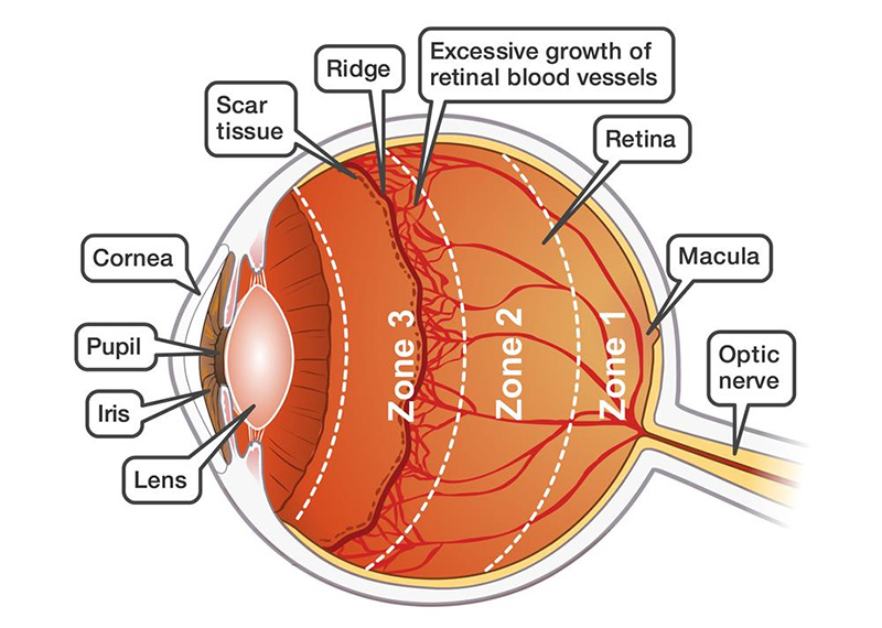
Retinopathy of prematurity (ROP) is a condition associated with  abnormal retinal vascular development in babies born at or before 30 weeks of gestation.

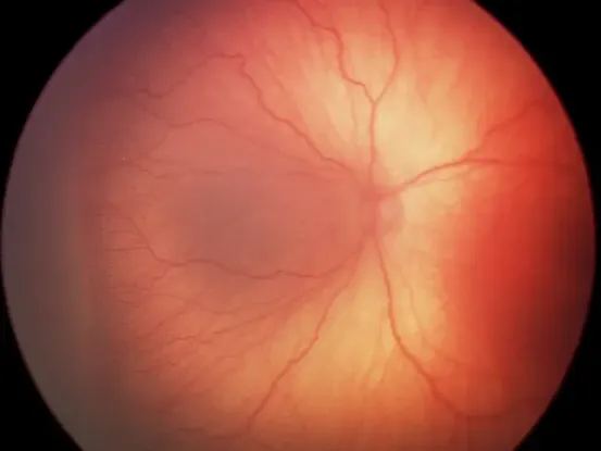
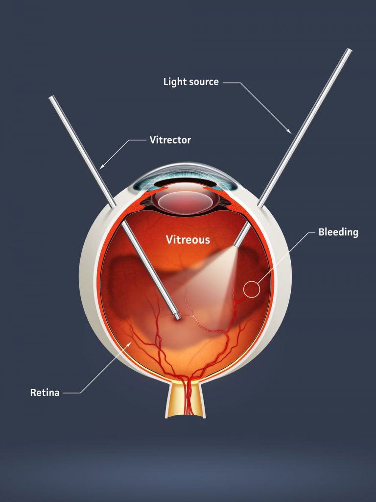
Advanced stages of ROP may require treatment, although this is not always the case. Milder stages are very common, and most affected newborns will recover completely. However, if advanced cases remain untreated, it can lead to blindness.

The most effective method of detecting ROP in premature infants is through regular screening, and this should continue until the retina has fully vascularised.

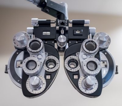
Babies who are born prematurely or those who develop any stage of ROP can develop other eye conditions like strabismus, amblyopia or refractive errors later in life. Therefore,  they should be assessed regularly by a paediatric ophthalmologist to monitor the potential development of these conditions after they turn one.

Chapter 1

What is ROP?

ROP is a disease in which abnormal blood vessels are formed in the immaturely developed retina of premature babies.

When the retina is developing normally, the blood vessels grow outwards from the optic disc to the periphery of the retina, usually between 20 weeks and full term.
retinopathy-of-prematurity

This can be disrupted in pre-term infants when vessel growth is temporarily slowed, resulting in increased production of vascular growth factors, such as VEGF (vascular endothelial growth factor). This stimulated vessel growth can be excessive, causing abnormal fibro-vascular tissue to proliferate anterior to the retina. If this abnormal tissue is not detected and treated in a timely manner, it can cause traction over the retina, leading to retinal detachment and permanent visual impairment.

Chapter 2

What causes Retinopathy of Prematurity?

While gestational age is the most important risk factor leading to development of ROP (according to the ANZNN 2013 Report, more than 50 per cent of babies born after less than 27 weeks’ gestation have some degree of ROP), there are other risk factors associated with the onset of the condition.
What causes Retinopathy of Prematurity

These can include:

  • Birth weight
  • Hyperopia and/or fluctuating oxygen tension
  • Fetal growth restriction
  • Sepsis
  • Limited postnatal growth
  • Birth weight is also an important risk factor, as smaller, preterm babies (particularly those weighing under 1250 g) were found to be more likely to develop ROP.
Chapter 3

The grading of ROP

Zone : Zone defines the extent of vascularisation.

Zone 1: Refers to a circle with a radius that is twice the distance from the centre of the optic disc to the macula.

Zone 2: Refers to a circle with a radius the same distance from the centre of the optic disc to the nasal margin of the retina (ora serrata).

Zone 3: Refers to the remainder of the retina, a crescent-shaped zone that largely involves temporal retina.
The grading of ROP

Stage: Stage defines the severity of the disease.

Stage 1: There is a thin but clear white line that separates the avascular retina anteriorly from the posteriorly vascularised retina.

Stage 2 – There is a ridge that arises out of the demarcation line and extends above the retina. It will either be white or pink.

Stage 3: Extraretinal fibrovascular proliferation or neovascularisation extends into the vitreous from the ridge.

Stage 4: Retinal detachments, which are generally concave and circumferential. They are further classified into 4A (extrafoveal) and 4B (foveal) categories.

Stage 5: Retinal detachments are usually tractional but can sometimes be exudative. They are usually funnel-shaped.

Plus: Plus disease reflects the activity of the ROP, such as increased venous dilatation and arteriolar tortuosity of the posterior retinal vessels. Iris vascular engorgement, poor pupillary dilatation (rigid pupil) and vitreous haze are associated changes. To be characterised as Plus disease, two quadrants of the retina must be affected,

Pre-plus disease: Pre-plus disease is when with the retinal vessel tortuosity and dilatation is not sufficiently abnormal to reach the criteria of Plus disease, but is nevertheless greater than that regarded as normal.

Chapter 4

How is ROP diagnosed in premature babies?

ROP has no signs or symptoms, when it first develops in premature babies. The only way to detect it, is with regular examinations and screening by a specialist paediatric ophthalmologist.

Generally, babies born before 32 weeks of gestation and those weighing less than 1250 g should regularly be examined and assessed by a paediatric ophthalmologist, with the precise timing of a first examination depending on their gestational age at birth. The timing of future examinations should subsequently be determined by presence or absence of ROP and the zone and stage of the disease.

The examinations are regularly carried on until the retina is fully vascularised into zone 3, without active ROP. This can usually take upto 37 weeks of gestation.
How is ROP diagnosed in premature babies
Chapter 5

How is ROP treated?

For advanced stages of ROP (Stage 3 and 4) , laser treatment is the most effective form of treatment. The aim is to photo-coagulate the peripheral avascular retina, which in turn reduces the production of VeGF, that stimulatesvascular proliferation. Intervention with laser treatment can prevent retinal detachment and therefore prevent blindness.

ROP can also ber treated with injections of VEGF inhibitors in the eye designed to reduce the production of VeGF thereby reducing the abnormal vascular proliferation. These injections can be administered in conjunction with or as an alternative to laser treatment in babies who are too sick to go ahead with laser treatment.

If a case of ROP is advanced with retinal detachment, retinal detachment surgery can be attempted. These surgeries may involve either repositioning the retina by using a scleral buckle around the eye or by performing vitrectomy and repositioning the retina through the inside of the eye.
How is ROP treated
Chapter 6

Ongoing follow up and care of preterm infants

In general, children born prematurely are at a risk of developing eye problems throughout their formative years. These can include refractive errors, strabismus and amblyopia.

Therefore, regular examination and assessment by a paediatric ophthalmologist is important to identify and treat any potential visual or eye concerns in a timely manner.

If you’re looking for an efficient way to detect and treat retinopathy of prematurity in Adelaide, we recommend booking an appointment with Dr Swati Sinkar. Dr. Swati is the first Paediatric Ophthalmologist to start Retinopathy of Prematurity screening service in Northern Adelaide Local Health Network, SA 

Related articles.

Strabismus (Squint Eyes)- All You Need to Know

November 14, 2021 | Eye Conditions

Strabismus, more commonly known as squint or crossed eyes, is the term used when a person’s eyes do not point in the ...

Read More

Eyelid Malposition – Causes, Surgery & Treatment

October 30, 2021 | Eye Conditions

There’s no doubt that our eyes are one of the most defining features of our physical appearance. Due to this, for ove...

Read More

What is Digital Eye Strain and How Can You Prevent It?

October 10, 2021 | Eye Conditions

There is no question that the COVID-19 pandemic has impacted different aspects of our health and wellbeing. This incl...

Read More